Scoliosis Screening
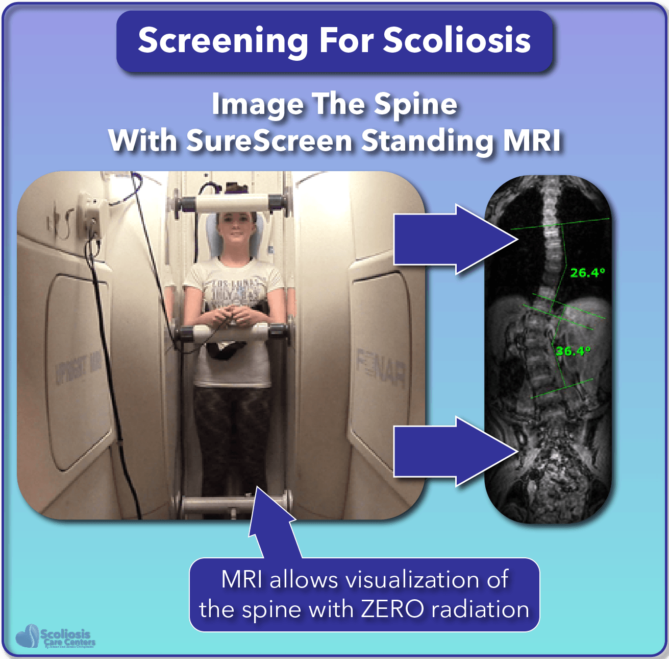
ScoliSnap Screen Standing MRI
Stop Using Outdated and Inaccurate Screening Methods
Screening children for scoliosis using visual methods can easily miss a curve, or suggest there is a curve when there isn’t one at all. While x-ray can detect a curve, it uses harmful ionizing radiation and should be avoided whenever possible.

The revolutionary ScoliSnap Screen Scoliosis MRI Screening provides an accurate, 100% radiation-free way of looking at the spine and detecting scoliosis. Scoliosis screening is now a sure thing compared to the inaccurate methods of the past. If you or your child have already been diagnosed with scoliosis, you can find information about scoliosis monitoring using MRI here .
Benefits of ScoliSnap MRI Screening for Scoliosis
- NEVER MISS A CURVE: ScoliSnap Screen uses standing MRI to actually look at the spine instead of simply looking at the surface of the back or checking for rib humps.
- EARLY DETECTION: Science shows the earlier a curve is caught and treated, the better the result1. ScoliSnap Screen allows a curve to be detected when it first appears, allowing early intervention and a 100% success rate for preventing surgery when treated using the Silicon Valley Method.
- RADIATION-FREE: ScoliSnap Screen is 100% radiation free in contrast to x-ray which uses ionizing radiation.
- FAST: ScoliSnap Screen can be performed in just one (1) minute, making it as fast and easy as a visual method while being a sure-fire method of detecting a curve.
📅 Schedule Your ScoliSnap Screening Today
Secure your spot for our upcoming scoliosis screening event, spots are limited.
Schedule Your Screening NowOld Screening Method VS ScoliSnap Screen
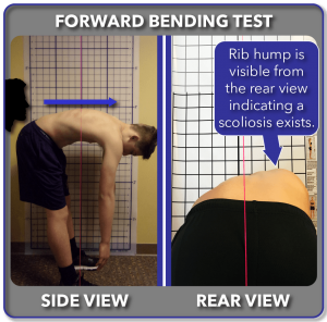
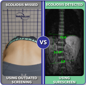
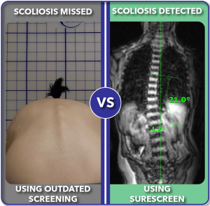
The traditional method of detecting scoliosis is to have a child bend forward while standing behind them to see if a rib hump is present. This method has been referred to as the “too late test” as it typically detects curves after they have already grown large in size.
The forward bending test will miss many curves when they are small. Here are real examples of real patients doing the forward bending test on the left and using ScoliSnap Screen on the right. Even when the forward bending test looks normal, ScoliSnap Screen can still detect the curve.

ScoliSnap Screen Options
ScoliSnap Scoliosis Screening
ScoliSnap Screen standing MRI scoliosis screening provides a quick and easy way to detect if a curve is present and how severe it is.
What is the ScoliSnap Screen Plus+ Clinical Exam?
Looks For Root Causes of Scoliosis
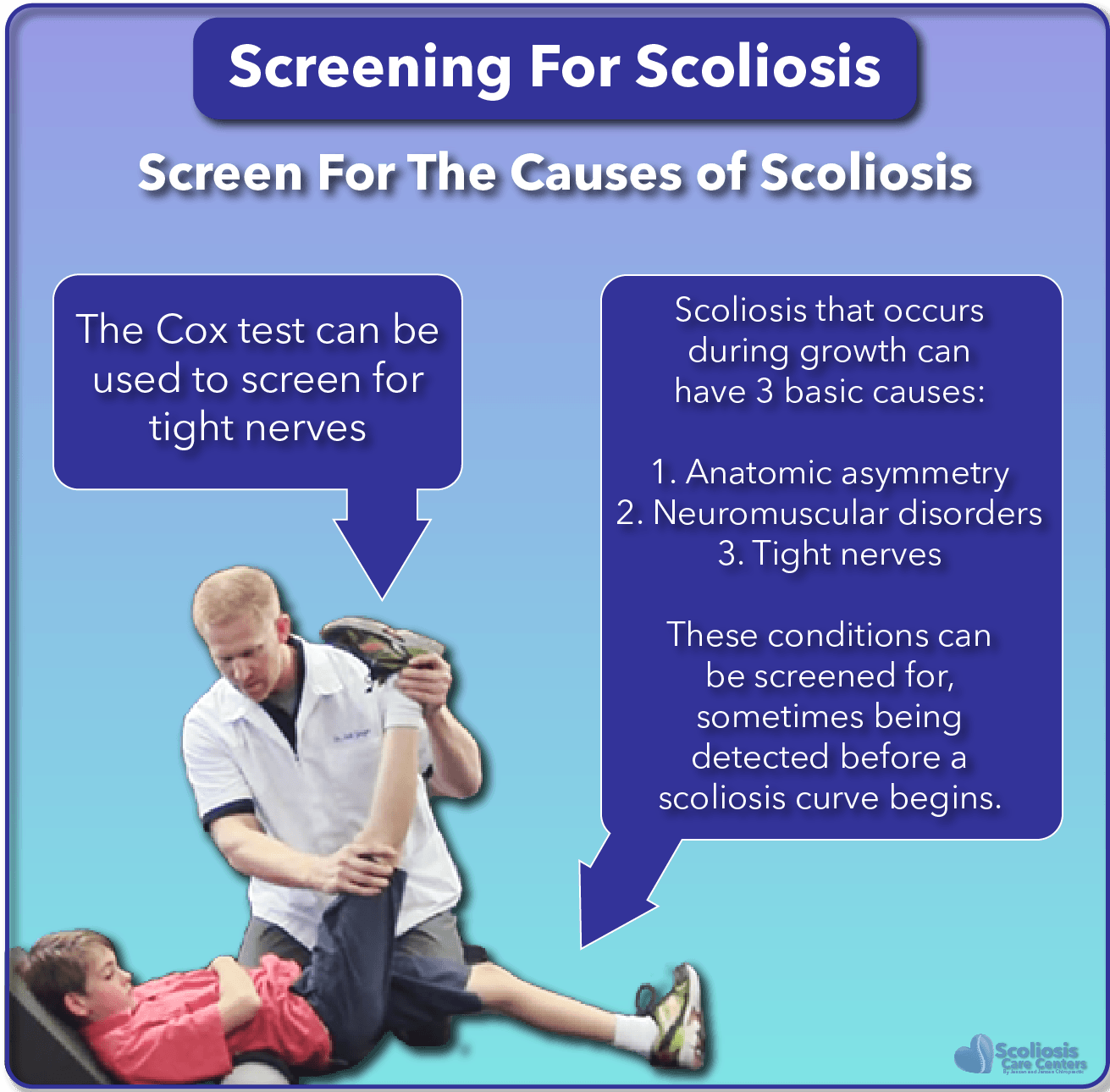
ScoliSnap Screen Plus+ provides the same accurate standing MRI scoliosis screen as the ScoliSnap Screen but also includes a clinical examination to see if any other underlying problems are present which can cause scoliosis to develop. This includes a tight spinal cord and nerve tension detected using the Cox test.
ScoliSnap Screen+ Uses More Accurate Traditional Screening Methods
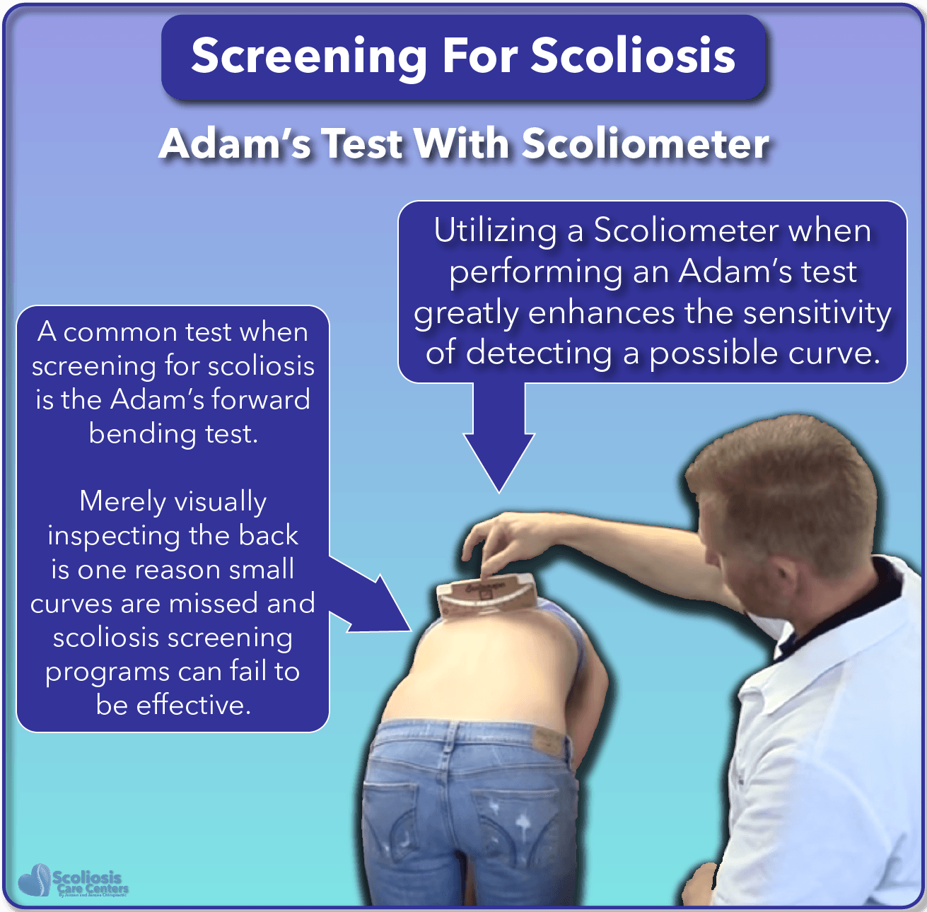
The ScoliSnap Screen Plus+ clinical exam uses traditional scoliosis screening methods such as the Adam’s test shown to the right, but with improved accuracy—tools like the Scoliometer make these tests far more sensitive.
Uses More Accurate Traditional Tests
The ScoliSnap Screen Plus+ clinical exam uses traditional scoliosis screening methods such as the Adam’s test shown below, but with improved accuracy. By using additional tools such as the Scoliometer, even traditional screening methods can be improved to be more accurate.
Non-traditional Risk Assessments
Studies have shown that symptoms such as joint hypermobility can be associated with scoliosis 2 and risk for surgery 3. Based on this scientific and medical research, the ScoliSnap Screen Plus+ clinical exam screens for ligament hypermobility; a tight spinal cord; and other warning signs of a potential scoliosis so that problems are detected early and allow early treatment and success.

References
- Shakil, Halima; Iqbal, Zaheen A.; Al-Ghadir, Ahmad H. (2014): Scoliosis. Review of types of curves, etiological theories and conservative treatment . Journal of Back and Musculoskeletal Rehabilitation 27(2), 111–115. DOI: 10.3233/BMR-130438.
- Czaprowski, Dariusz; Kotwicki, Tomasz; Pawłowska, Paulina; Stoliński, Lukasz (2011): Joint hypermobility in children with idiopathic scoliosis. SOSORT award 2011 winner . Scoliosis 6:22. DOI: 10.1186/1748-7161-6-22.
- Haller, Gabe; Zabriskie, Hannah; Spehar, Shelby; et al. (2018): Lack of joint hypermobility increases the risk of surgery in adolescent idiopathic scoliosis . Journal of Pediatric Orthopedics Part B 27(2), 152–158. DOI: 10.1097/BPB.0000000000000489.
📅 Schedule Your ScoliSnap Screening Today
Secure your spot for our upcoming scoliosis screening event, spots are limited.
Schedule Your Screening Now