Standing MRI Isn’t Advanced — It’s the Minimum Standard for Proper Scoliosis Care
At Scoliosis Care Centers, we rely on 100% radiation-free standing MRI—rather than traditional X-rays—to safely and accurately monitor treatment progress and Cobb angles.
Radiation-free. Real-time. Remarkably more accurate.
Traditional scoliosis monitoring relies heavily on X-rays—despite the risks of repeated radiation exposure and long wait times between scans. At Scoliosis Care Centers™, we use 100% radiation-free standing MRI instead. It’s not just safer — it’s smarter.
This kind of monitoring shouldn’t be rare—it should be the standard of care.
Why Standing MRI Makes All the Difference in Your Care
Standing MRI Every 3 Months to Monitor Progress Safely
Why Frequent Monitoring MattersIn-Brace MRI Monitoring to Ensure Continued Correction
See How We Track Brace PerformanceTailoring Treatment Plans Through Real-Time MRI Insights
How MRI Improves Brace DesignWhy Frequent MRI Monitoring Matters
Most scoliosis monitoring uses X-rays every 6–12 months. But for growing kids, that’s too far apart. Curves can worsen quickly during a growth spurt. Limiting imaging to once or twice a year—just to minimize radiation—can allow scoliosis to progress unchecked.
That’s why Scoliosis Care Centers™ uses 100% radiation-free standing MRI every three months. We take these scans while the patient is standing and not wearing the brace. This shows us how the spine is truly responding to treatment. Cobb angle measurements are plotted over time, so families can see progress clearly—and intervene early if needed.
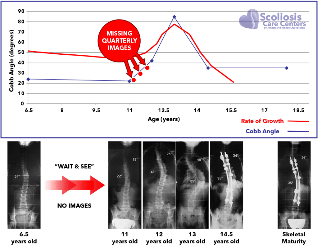
This 11-year-old’s curve worsened from 22° to 42° in one year.
With quarterly MRI monitoring (shown by red arrows), this progression could have been caught and corrected before reaching surgical range.
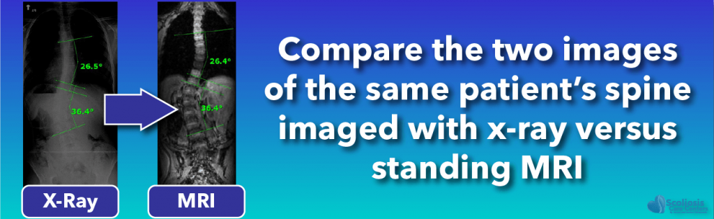
MRI vs. X-Ray: MRI provides clearer, more frequent insights—without exposing the spine to harmful radiation.
How We Track Brace Performance with MRI
The Silicon Valley Brace™ is one of the few scoliosis braces designed to be fully MRI-compatible. This allows us to conduct standing MRI scans every three months—while the patient is wearing the brace—to evaluate its effectiveness in real time. No radiation. No guessing.
As children grow, their bodies change—spinal alignment improves, body shape shifts, and brace fit naturally becomes less precise. That’s why frequent in-brace MRI monitoring is critical. It helps us detect subtle performance issues early, make targeted adjustments, and ensure the brace continues to guide the spine toward straighter alignment.

Real-time feedback without radiation.
MRI shows how well the brace is correcting the spine—so adjustments can be made immediately, not months later.
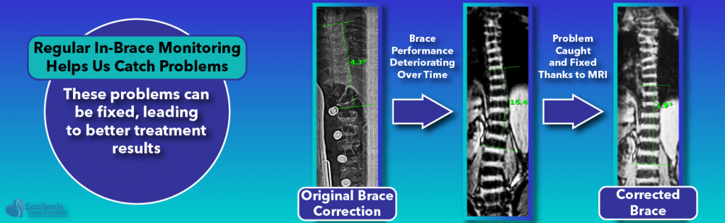
Don’t wait for problems to show up on an X-ray.
MRI helps us detect brace fit issues as they arise—keeping treatment on track.
How MRI Feedback Improves Brace Design
At Scoliosis Care Centers™, even the design process for our Silicon Valley Brace™ is guided by standing MRI. As soon as the brace is first fitted, we perform a real-time MRI scan with the patient wearing the brace. This allows us to measure exactly how the spine is responding—and make any necessary refinements immediately.
MRI is completely radiation-free. This means we can repeat the process often to perfect the brace’s performance. Instead of waiting months for X-ray feedback or relying on guesswork, we use real-time imaging to ensure each brace delivers maximum in-brace correction from day one. We apply the same MRI-guided approach to evaluate other scoliosis treatments as well.

Better feedback leads to better results.
Standing MRI gives us immediate insight into how each brace is performing—and how it can be improved.
What If MRI Isn’t an Option?
In rare cases where standing MRI is not possible, we use low-dose, highly-shielded X-ray imaging to minimize radiation exposure. Unlike many clinics, we carefully track the number of X-rays each patient receives to ensure total exposure stays as low as possible—without sacrificing the quality of care.

When X-rays are necessary, we do them the right way.
We use shielding, dose reduction protocols, and careful tracking to protect your child’s long-term health.
Don’t Settle for Outdated Scoliosis Monitoring
Standing MRI allows us to track, adjust, and improve scoliosis treatment without exposing patients to harmful radiation. If your child is still being monitored by X-ray alone, it’s time for a better standard of care.
Schedule a Free Consultation